Ultrasound-assisted neuraxial blockade
A four-step scan of the lumbar spine: find the L3/4 space, confirm you are in the midline, then measure the depth and angle to the ligamentum flavum before the needle goes anywhere near the patient.
- 1 Position the patient
Position the patient as you would for the block itself — sitting or lateral, back flexed, shoulders relaxed forward — and keep them in that position for both the scan and the block. Use a low-frequency curvilinear probe with an abdominal preset and a starting depth of around 8 cm.
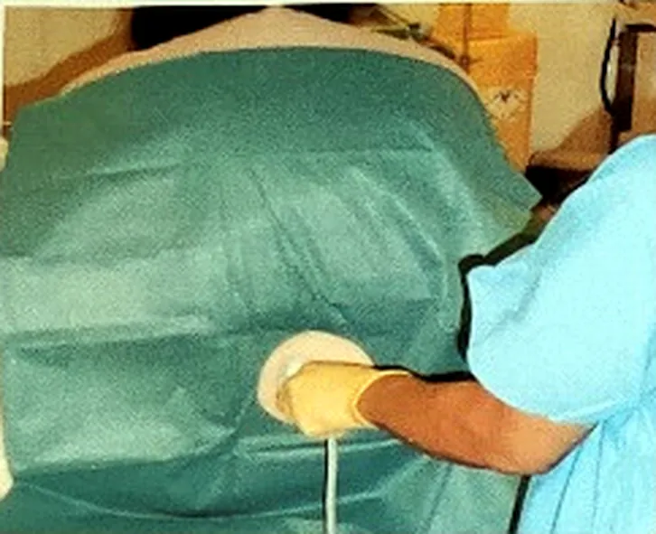
Scan and block in the same position. - 2 Longitudinal paramedian scan
With the probe in the paramedian longitudinal plane, scan upwards from the sacrum, which is seen as a series of continuous bumps forming a hyperechoic line. Identifying the laminae of the lumbar vertebrae lets you correctly identify the L3/4 space. It also confirms you are following the midline, which negates problems from anatomical anomalies such as scoliosis.
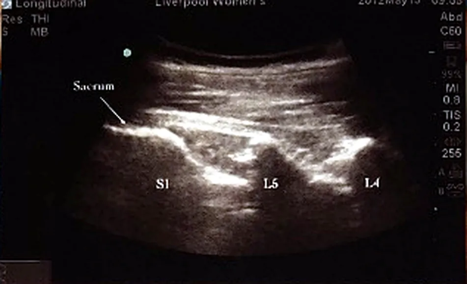
Sacrum, then S1, L5 and L4 counted upwards. - 3 Horizontal scan
Having identified the desired space, rotate the probe through 90°, maintaining skin contact throughout, to give a transverse image. When the probe is level with the spinous process, the process is seen as a superficial hyperechoic structure casting a dark shadow below it.
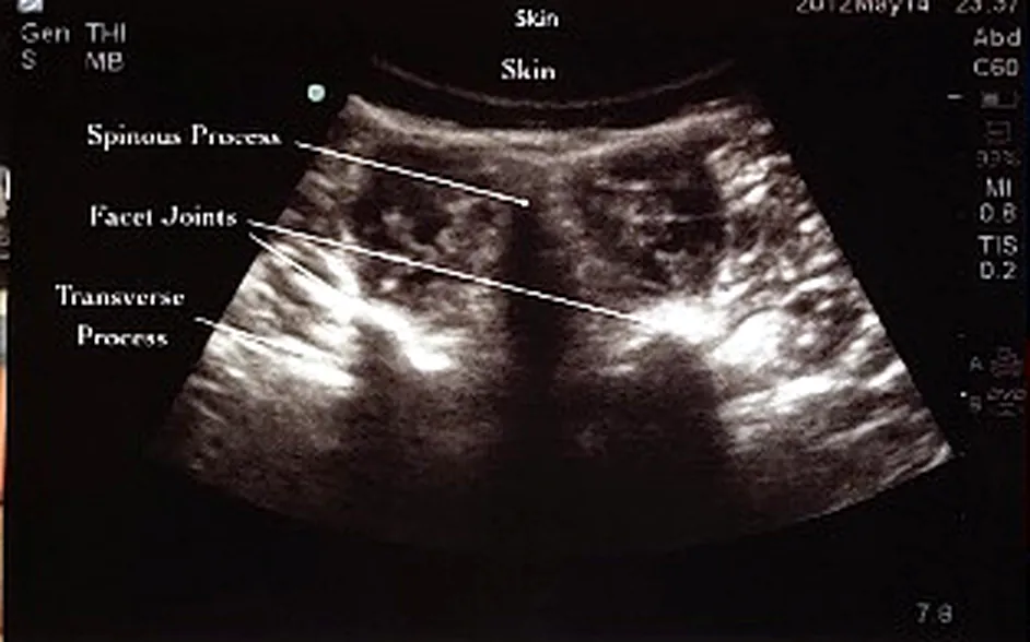
Spinous process, facet joints and transverse processes in transverse section. - 4 Identify the space
Move the probe superiorly or inferiorly from the spinous process to locate the inter-space. Staying correctly in the midline gives a symmetrical image, bordered by the articular processes.
The transverse picture allows identification of several structures, most importantly the ligamentum flavum. Using the calipers on the ultrasound machine, measure the distance from skin to ligament. Note the angle at which the ligamentum flavum is seen, and replicate that angle on needle insertion.
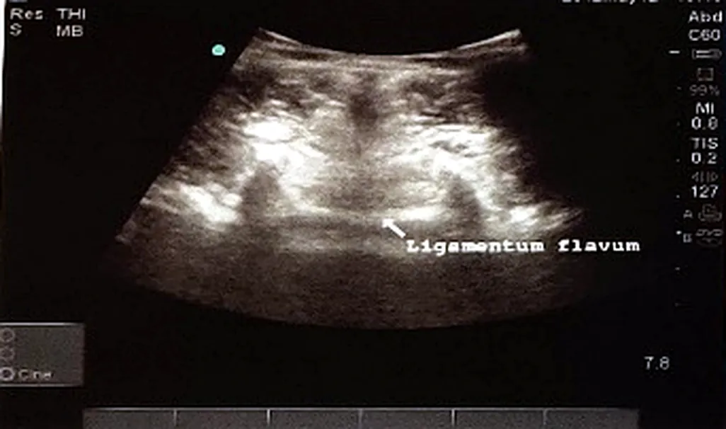
Symmetrical inter-space view with the ligamentum flavum labelled.
Both the measured skin-to-ligament depth and the angle of insonation are worth writing down before you scrub — the depth tells you when to expect loss of resistance, and the angle is the one you reproduce with the needle.
Adapted from the departmental poster Ultrasound assisted Neuraxial blockade: A Step by Step Guide by Dr A Ssenoga, Dr C Chevannes, Dr A Bhalla, Dr P Barclay and Dr H McNamara, Liverpool Women's Hospital.